Answer
to Case 24
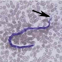
The microfilaria in the images is that of Loa loa (African eye worm).
Diagnostic features to distinguish this microfilaria from other species
included:
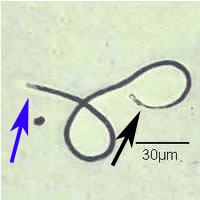
- Size (230 to 250 micrometers in length by 6 to 8.5 micrometers in diameter in stained blood films, 270 to 300 micrometers in length in formalin preparations),
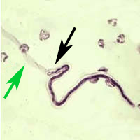
- A tapered tail with nuclei extending to the tip (black arrows),
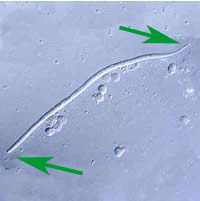
- The presence of a sheath (green arrows) normally seen on hematoxylin stained blood films or using differential interference contrast (DIC) for unstained, wet mounts (Figure D below). The sheath is usually not visible in Giemsa stained blood films, though a halo or clear space (Figure B below, blue arrow) can sometimes be seen and can be interpreted as a sheath being present.
A note of caution: Microfilariae undergo varied degrees of shrinkage in stained blood films, and as a consequence, measurement of microfilaria length in stained blood films is helpful, but it must be remembered that individual microfilariae will often measure appreciably smaller than stated sizes, especially in thin blood films. Many times infection with Loa tend to be asymptomatic, however, the adult worms can wander widely in the human body. This is especially troubling when the worms cross the conjunctiva of the eye. The microfilaria of Loa loa can be found in peripheral blood and are most numerous during mid-day samples.
 |
 |
| A | B |
 |
 |
| C | D |