Answer
to Case 35
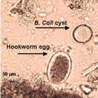
Two parasites were present in this case: hookworm eggs (most likely Necator
americanus based on geographical location) and Balantidium coli
(cysts and trophozoites). Hookworm eggs are fairly easy to identify
from their general appearance, size (55 to 65 micrometers by 36 to 40
micrometers), and thin shells. The embryonated egg (Figure E)
may have been tricky since Strongyloides stercoralis eggs, although
usually not seen in stool preparations, are similar in appearance.
Occasionally, one may see embryonated hookworm eggs if the specimen
was kept at room temperature, even for a short period of time, before
being preserved in formalin. A mixture of fully embryonated and
partially embryonated eggs suggests hookworm infection. If this
had been an infection with S. stercoralis, one would expect to
see larvae instead of eggs, but, if eggs were present, they would all
most likely be fully embryonated. We did not see any nematode larvae
in this specimen that might suggest an infection with S. stercoralis.
Morphologically, eggs of hookworms and S. stercoralis are difficult
to distinguish when embryonated.
The images of the cysts of B. coli (from a wet mount made from the concentrate of this specimen) were difficult to diagnose. Sometimes, especially in older cysts, there is not much morphological detail to be seen other than size (about 50 to 70 micrometers). In the images (Figure C), one can see the cyst wall and cytoplasmic inclusions instead of structures such as nuclei that would normally be seen in ameba cysts. Cysts of B. coli may have some or none of the following diagnostic features: evidence of cilia inside the cyst wall, a large macronucleus, a smaller micronucleus, and cytoplasmic inclusions or vacuoles. We have added arrows to the original images sent in this case to highlight some of these features.
 |
| A |