Answer to
Case 48
This was an artifact that resembled a nematode larva. The artifact was
initially considered to be a first-stage Strongyloides stercoralis larva. Several morphologic features indicated this was not a
Strongyloides larva. The
size and general shape of the structure was not consistent with a parasitic
nematode larva. First-stage (rhabditiform) Strongyloides
larvae measure 200 to 400 micrometers in length by 15 to 20 micrometers
in diameter, while third-stage (infective, filariform) larvae measure
500 to 600 micrometers in length by 15 to 16 micrometers in diameter.
This object was slightly longer than the maximum for first-stage larvae
and shorter than third-stage larvae. Additionally, the diameter
was considerably larger than Strongyloides larvae of either stage.
First-stage larvae of Strongyloides have a long, tapered tail,
whereas this object had a very short blunt end by comparison. Although
some very small, fine particulate matter is visible in the object, it
does not resemble typical nuclei seen in stained nematode larvae, and
the object appears to be homogenous in appearance and lacks any internal
structure.
A number of people thought the object was Ascaris larva, and several suggested Necator. No one suggested Toxocara, but this should have been considered as it is a more likely occurrence than Necator (or other hookworm larvae). All of these parasitic nematode larvae can be excluded for many of the same reasons as used to rule out Strongyloides. The object in question is larger, especially in diameter, than Toxocara or hookworm, both of which are no more than about 21 micrometers. Second stage Ascaris larvae are also much smaller (approximately 300 by 15 micrometers). Third stage Ascaris larvae can reach 1.6 millimeters in length by 26 to 50 micrometers in diameter. In addition to size differences, the two most helpful features in distinguishing an artifact from a nematode larva is the absence of recognizable internal structure and the very truncated, blunt end that does not resemble a tail on a larva.
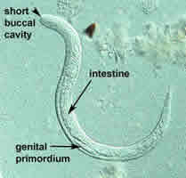
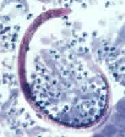
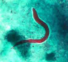
Several images of Strongyloides larvae are shown below for comparison to the artifact. Figure A below is from a wet mount of stool with a first-stage larva showing the general larval features (captured using DIC); Figure B below shows a stained filariform larva found in a sputum sample; and Figure C below illustrates a rhabditiform (first-stage) larva in a trichrome stained stool smear. Although few internal features are evident in these stained larvae, the general shape of the larvae is evident, and clearly distinct from the artifact presented in this case. One must remember that in disseminated Strongyloides cases, in addition to both first and third-stage larvae, adult female worms, or even eggs, might also be found.
Click here for more information about Strongyloides.
 |
 |
| A | B |
 |
| C |