|
|
[Last Modified: ] |
Case
55
A 33-year-old man returned to the United States after spending four years
in Africa, mostly Kenya, working as an agricultural consultant.
He had several parasitic diseases during his four years in Africa.
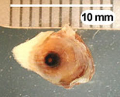
After returning to the United States, he noticed a painful, swollen lesion
with a black center on the top of his right foot. Initial diagnosis
from a health care provider was that the lesion was most likely a plantar
wart. The man did not agree with the physician and obtained a second
opinion from a health care provider who suggested that the lesion be biopsied.
The physician surgically removed the affected area and examined the
excised tissue. The physician saw what appeared to be eggs in the
tissue. The entire specimen was sent CDC's reference laboratory for identification
of the eggs. The man did not have any complications from the surgery
and the area healed within a few days. Figure A shows the
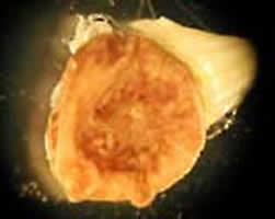
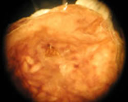
entire specimen; all other images were taken from the cut surface of the
tissue. Figures B and C were taken with a dissecting
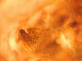
microscope equipped with adjustable zoom magnification. Figure D
was taken with a compound microscope using a 5× objective and a fiber-optic
light source to provide reflected light. What is your diagnosis?
Based on what criteria?
 |
 |
| A | B |
 |
 |
| C | D |
Click here for the answer to Case 55.
Case
56
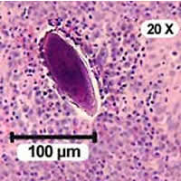
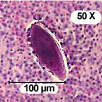
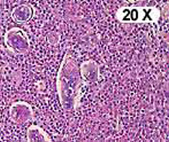
A 25-year-old man came to the United States from Ghana in 1997 and later
joined the U.S. Army. While in Ghana, the man bathed, swam, did
laundry, and drank from the Volta River, which was close to his home.
In early 2000, he began noticing blood in his urine. He was diagnosed
with a venereal disease, treated, and released. He continued to
have symptoms (hematuria) and was seen by a urologist, who ordered a bladder
biopsy. Figures A, B, C, and D show what was
found in H & E (hematoxylin and eosin) stained sections of his bladder wall. What is
your diagnosis? Based on what criteria?
 |
 |
| A | B |
 |
 |
| C | D |
Acknowledgement: This case was kindly provided by the U.S. Army.
Click here for the answer to Case 56.
|
|||