|
|
[Last Modified: ] |
Case
57
A 32-year-old woman from Sudan submitted a stool specimen as part of a general
physical examination. She was a refugee from Sudan and had been
living in a holding camp in the Middle East before relocating to Texas.
The woman’s health was compromised because of poor living conditions,
including inadequate sanitary facilities and exposure to animals.
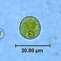
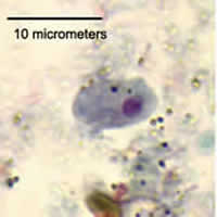
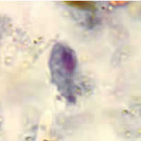
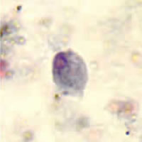
The parasitology lab at the Texas Department of Health, Bureau of Laboratories
identified the organism shown in Figures A and B. Figure
A was captured from a direct wet mount prepared from a formalin
preserved specimen and stained with Lugol’s iodine. Figure B,
which simulates focusing through four different planes, is a series
of four individual images of the same organism from a PVA stool smear
stained with the trichrome stain. The objects ranged in size from
10 to 18 micrometers in diameter. What is your diagnosis?
Based on what criteria?
 |
| A |
 |
| B (Simulating focusing) |
Acknowledgement: This case was kindly contributed by the Texas Department of Health, Bureau of Laboratories.
Click here for the answer to Case 57.
Case
58
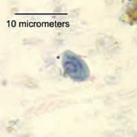
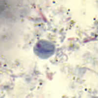
Laboratorians in the Division of Parasitic Diseases at CDC examined a stool specimen
from an HIV-positive patient. Examination of the stool specimen
included a formalin-ethyl acetate (FEA) concentration and smears stained
with trichrome. Figures A, B, and C show cysts
and Figures D, E, and F show trophozoites that were
found on the trichrome stained smear. What is your diagnosis?
Based on what criteria?
 |
 |
| A | B |
 |
 |
| C | D |
 |
 |
| E | F |
Click here for the answer to Case 58.
|
|||