|
|
[Last Modified: ] |
Case
9
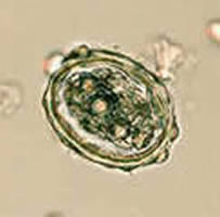
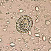
A 36-year-old woman with a history of travel to Puerto Rico submitted a
routine stool specimen. The images below were captured during wet
mount examination of the concentrate from a stool specimen. The images
are not on the same scale (see below). What is your diagnosis? Based on
what criteria?
 |
 |
| A | B |
A: 60 µm by 49 µm; B: 71 µm by 44 µm
 |
 |
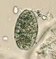
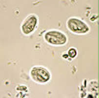
| C | D |
C: 12 µm by 14 µm; D: 55 µm by 27 µm
 |
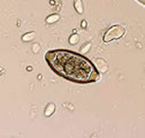
| E |
E: 50 µm by 44 µm
Acknowledgement: The images were kindly shared by Dr. Ray Kaplan, Atlanta.
Click here for the answer to Case 9.
Case
10
The Wisconsin State Laboratory of Hygiene (Tim Monson and Linda Kelly) sent
this image to CDC, with the following message:
“We are wondering if you can help us diagnose/rule out a body consistent with a Fasciola/Fasciolopsis egg. It measures around 140 micrometers in length and there is no apparent operculum present (not that we can detect anyway). Two stools were submitted in 10% formalin, and we could only find the one “egg” in question here. The patient history that we obtained is as follows: 26 year old male; travel to Cancun, Mexico 3/6 through 3/13; onset of symptoms on 3/13 (stomach pain, diarrhea [4/day], intestinal discomfort); stools collected on 3/18; symptoms resolved by 3/26.”
Is this a parasite egg? Based on what criteria? If not a parasite egg, what is it? Based on what criteria? What other examination(s) would you recommend?
Our colleagues from Wisconsin added: “The image was taken in black and white… We just used a Polaroid camera that fits onto one of the scope’s oculars.”
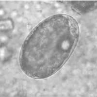 |
| A |
Click here for the answer to Case 10.
|
|||