|
|
[Last Modified: ] |
Case
67
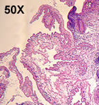
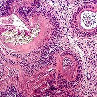
A 67-year-old woman visited her health care provider because of a subcutaneous
nodule located in the posterior area of her right shoulder. The
cyst was surgically removed. It measured three to four centimeters
in diameter and was filled with clear fluid. The material was submitted
for histological examination. Sections were made and stained with
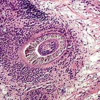
H & E (hematoxylin and eosin). Figure A shows a large
area of the stained section captured using 50× magnification (5× objective)
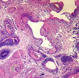
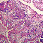
from one slide, and Figures B and C show cropped areas using
200× magnification (20× objective). What is your diagnosis?
Based on what criteria?
|
|||||||||
| A |
 |
 |
| B | C |
Click here for the answer to Case 67.
Case
68
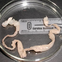
An adult man passed a segmented worm and submitted it to his local state health department
for identification. The worm was placed in 10% formalin and sent
to CDC (Figure A). In addition to gross examination,
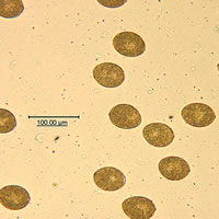
a segment was removed from the distal end and compressed between two glass
slides, producing the objects shown in Figure B. What is
your diagnosis? Based on what criteria?
 |
 |
| A | B |
Click here for the answer to Case 68.
Images presented in the monthly case studies are from specimens submitted for diagnosis or archiving. On rare occasions, clinical histories given may be partly fictitious.
|
|||