|
|
[Last Modified: ] |
|
|
|
| [Balantidium coli] |
|
|
|
|
|
|
|
|
Microscopy
 |
 |
 |
| A | B | C |
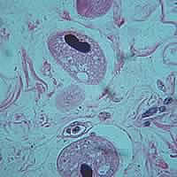
A:
Balantidium coli trophozoites in tissue, hematoxylin and eosin stain.
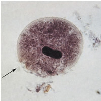
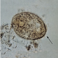
B, C: Balantidium coli
trophozoites. These are characterized by:
- their large size (40 µm to more than 70 µm).
- the presence of cilia on the cell surface, particularly visible in (C).
- a cytostome (arrows).
- a bean shaped macronucleus which is often visible, see (B), and a smaller, less conspicuous micronucleus.
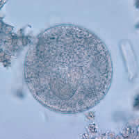 | D |
D: Balantidium coli cyst, unstained.
|
|||