|
|
[Last Modified: ] |
|
|
|
| [Cyclospora cayetanensis] |
|
|
|
|
|
|
|
|
Microscopy
It is recommended that
concentration procedures be used prior to microscopic examination. The sediment can be
examined using various techniques:
Wet mounts:
In wet mounts, Cyclospora oocysts appear as round organisms, 8 to 10
µm in diameter,
with a distinct oocyst wall (Images D, E). This
contrasts with oocysts of the two most frequent coccidian parasites infecting humans: Cryptosporidium
parvum and C. hominis oocysts are round but half the size, 4 to 6 µm in diameter (A,
B);
and Isospora belli oocysts are oblong and substantially larger (G,
H).
Microscopic examination of wet mounts can be enhanced by two techniques:
- UV fluorescence microscopy, where the oocysts autofluoresce under ultraviolet light (E). In comparison, Cryptosporidium sp. oocysts do not autofluoresce to any extent, while those of Isospora belli do (H). The walls of the oocysts fluoresce brightly, while their interiors do not, which differentiates Cyclospora from other objects of similar size and shape. Because this fluorescence is very evident, UV fluorescence microscopy is a sensitive technique for rapidly examining stool sediments.
- Differential interference contrast (DIC, Nomarsky), which shows Cyclospora oocysts as nonrefractile spheres that contain undifferentiated cytoplasm or refractile globules (D).
The two main disadvantages of wet mounts are that some laboratories do not have the equipment necessary for UV fluorescence microscopy or for DIC, and wet mount preparations cannot be archived as permanent records.
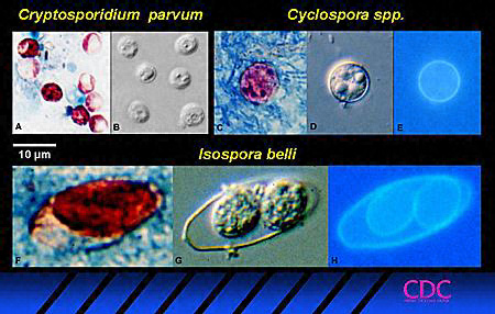 |
| Figures A - H |
Three coccidian parasites that most commonly infect humans, seen in acid-fast stained smears (A, C, F), bright-field differential interference contrast (B, D, G) and UV fluorescence (E, H).
Stained Smears:
Conventional parasitology stains do not reliably demonstrate Cyclospora oocysts.
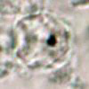 |
| I |
I: Cyclospora oocyst stained with trichrome; while the oocyst is visible, the staining characteristics are inadequate for a reliable diagnosis.
Two special stains allow a
more reliable diagnosis:
Modified
acid-fast stains are the standard for staining coccidian oocysts (Cyclospora spp.,
Cryptosporidium spp., Isospora spp.). In modified acid-fast stains, Cyclospora oocysts
retain the same size as in wet mounts, but frequently they are not perfectly round.
In
addition, the oocyst wall is less apparent and can take a wrinkled appearance, and appear
collapsed or distorted on one side. The oocysts are variably stained, with different
oocysts ranging from colorless to deep purple. This variability can lead to
misidentification and constitutes a drawback.
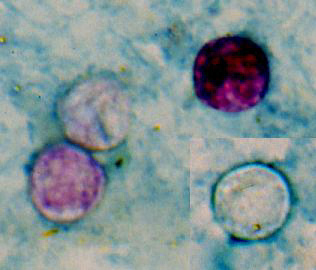 |
| J |
J: Four Cyclospora oocysts from fresh stool fixed in 10% formalin and stained with modified acid-fast stain. Compared to wet mount preparations, the oocysts are less perfectly round and have a wrinkled appearance. Most importantly, the staining is variable among the four oocysts.
A modified safranin stain (the modification consisting in heating in a microwave during staining) allows a uniform staining (red to reddish-orange) of the oocysts, whose wall appear wrinkled. This technique is not only more reliable than the modified acid-fast stain, but it is also rapid and easy to perform in most clinical laboratories.
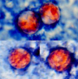 |
| K |
K: Four Cyclospora oocysts from fresh stool fixed in 10% formalin and stained with safranin, showing the uniform staining of oocysts by this method.
Reference:
Eberhard ML, Pieniazek NJ, Arrowood MJ. Laboratory diagnosis of Cyclospora infections. Arch Pathol Lab Med 1997;121:792-797.
|
|||