|
|
[Last Modified: ] |
|
|
|
| [Hymenolepis nana] [Hymenolepis diminuta] |
|
|
|
|
|
|
|
|
Causal Agents:
Hymenolepiasis is caused by two
cestodes (tapeworm) species, Hymenolepis nana (the dwarf tapeworm,
adults measuring 15 to 40 mm in length) and Hymenolepis dimnuta (rat
tapeworm, adults measuring 20 to 60 cm in length). Hymenolepis diminuta
is a cestode of rodents infrequently seen in humans and frequently found in
rodents.
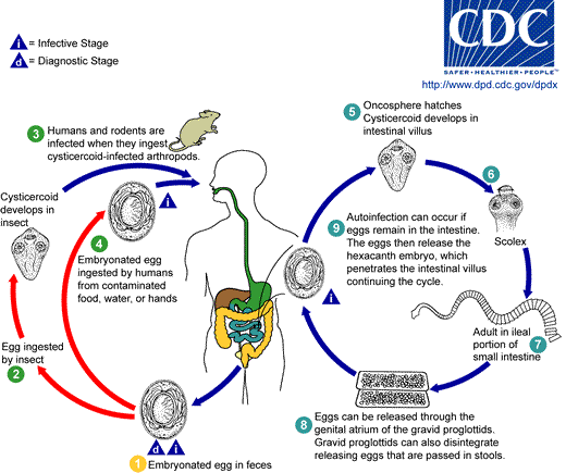
Eggs of
Hymenolepis nana are immediately infective when passed with the stool
and cannot survive more than 10 days in the external environment
![]() .
When eggs are ingested by an arthropod intermediate host
.
When eggs are ingested by an arthropod intermediate host
![]() (various species of beetles and fleas may serve as intermediate hosts),
they develop into cysticercoids, which can infect humans or rodents upon
ingestion
(various species of beetles and fleas may serve as intermediate hosts),
they develop into cysticercoids, which can infect humans or rodents upon
ingestion
![]() and develop into adults in the small intestine. A morphologically
identical variant, H. nana var. fraterna, infects rodents
and uses arthropods as intermediate hosts. When eggs are ingested
and develop into adults in the small intestine. A morphologically
identical variant, H. nana var. fraterna, infects rodents
and uses arthropods as intermediate hosts. When eggs are ingested
![]() (in contaminated food or water or from hands contaminated with feces), the
oncospheres contained in the eggs are released. The oncospheres (hexacanth
larvae) penetrate the intestinal villus and
develop into cysticercoid larvae
(in contaminated food or water or from hands contaminated with feces), the
oncospheres contained in the eggs are released. The oncospheres (hexacanth
larvae) penetrate the intestinal villus and
develop into cysticercoid larvae
![]() .
Upon rupture of the villus, the cysticercoids return to the intestinal
lumen, evaginate their scoleces
.
Upon rupture of the villus, the cysticercoids return to the intestinal
lumen, evaginate their scoleces
![]() ,
attach to the intestinal mucosa and develop into adults that reside
in the ileal portion of the small intestine producing gravid proglottids
,
attach to the intestinal mucosa and develop into adults that reside
in the ileal portion of the small intestine producing gravid proglottids
![]() .
Eggs are passed in the stool when released from proglottids through its
genital atrium or when proglottids disintegrate in the small intestine
.
Eggs are passed in the stool when released from proglottids through its
genital atrium or when proglottids disintegrate in the small intestine
![]() .
An alternate mode of infection consists of internal autoinfection, where
the eggs release their hexacanth embryo, which penetrates the villus
continuing the infective cycle without passage through the external
environment
.
An alternate mode of infection consists of internal autoinfection, where
the eggs release their hexacanth embryo, which penetrates the villus
continuing the infective cycle without passage through the external
environment
![]() .
The life span of adult worms is 4 to 6 weeks, but internal autoinfection
allows the infection to persist for years.
.
The life span of adult worms is 4 to 6 weeks, but internal autoinfection
allows the infection to persist for years.
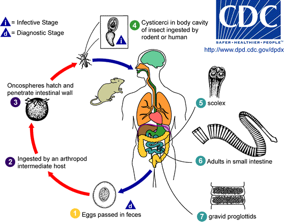
Eggs of Hymenolepis
diminuta are passed out in the feces of the infected definitive host
(rodents, man)
![]() .
The mature eggs are ingested by an intermediate host (various arthropod
adults or larvae)
.
The mature eggs are ingested by an intermediate host (various arthropod
adults or larvae)
![]() ,
and oncospheres are released from the eggs and penetrate the intestinal wall
of the host
,
and oncospheres are released from the eggs and penetrate the intestinal wall
of the host
![]() ,
which develop into cysticercoid larvae. Species from the genus Tribolium are
common intermediate hosts for H. diminuta. The cysticercoid larvae
persist through the arthropod's morphogenesis to adulthood. H. diminuta infection is acquired by
the mammalian host after ingestion of an intermediate host carrying the cysticercoid larvae
,
which develop into cysticercoid larvae. Species from the genus Tribolium are
common intermediate hosts for H. diminuta. The cysticercoid larvae
persist through the arthropod's morphogenesis to adulthood. H. diminuta infection is acquired by
the mammalian host after ingestion of an intermediate host carrying the cysticercoid larvae
![]() . Humans can be accidentally infected through the ingestion of
insects in precooked cereals, or other food items, and directly from the
environment (e.g., oral exploration of the environment by children).
After ingestion, the tissue of the infected arthropod is digested
releasing the cysticercoid larvae in the stomach and small intestine.
Eversion of the scoleces
. Humans can be accidentally infected through the ingestion of
insects in precooked cereals, or other food items, and directly from the
environment (e.g., oral exploration of the environment by children).
After ingestion, the tissue of the infected arthropod is digested
releasing the cysticercoid larvae in the stomach and small intestine.
Eversion of the scoleces
![]() occurs shortly after the cysticercoid larvae are
released. Using the four suckers on the scolex, the parasite attaches to the small intestine wall.
Maturation of the parasites occurs within 20 days and the adult worms can
reach an average of 30 cm in length
occurs shortly after the cysticercoid larvae are
released. Using the four suckers on the scolex, the parasite attaches to the small intestine wall.
Maturation of the parasites occurs within 20 days and the adult worms can
reach an average of 30 cm in length
![]() . Eggs are released in the small
intestine from gravid proglottids
. Eggs are released in the small
intestine from gravid proglottids
![]() that disintegrate after breaking off from
the adult worms. The eggs are expelled to the environment in the
mammalian host's feces
that disintegrate after breaking off from
the adult worms. The eggs are expelled to the environment in the
mammalian host's feces
![]() .
.
Geographic
Distribution:
Hymenolepis nana is the
most common cause of all cestode infections, and is encountered worldwide. In
temperate areas its incidence is higher in children and institutionalized groups. Hymenolepis
diminuta, while less frequent, has been reported from various areas of the world.
|
||||||||