|
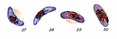
Plasmodium
falciparum: Gametocytes
Figs. 27, 28:
Mature macrogametocytes (female); Figs. 29, 30: Mature microgametocytes (male).
Illustration from:
Coatney GR, Collins WE, Warren M, Contacos PG. The Primate Malarias. Bethesda:
U.S. Department of Health, Education and Welfare; 1971.
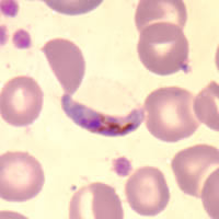
Smears from patients:
Plasmodium
falciparum gametocytes, when mature are crescent or sausage shape. The
red blood cell is often distorted or not visible.
 |
 |
| A |
B |
 |
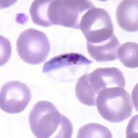 |
| C |
D |
A, B,
C, D: Gametocytes of P. falciparum in thin blood
smears. Note the presence of a “Laveran’s bib," which is not always
visible.
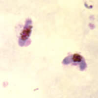 |
| E |
E: Two
gametocytes captured from a thick blood smear.
|