|
Plasmodium
falciparum: Blood Stage Parasites
Thin Blood Smears
Fig. 1: Normal
red cell; Figs. 2-18: Trophozoites (among these, Figs. 2-10
correspond to ring-stage trophozoites); Figs. 19-26: Schizonts (Fig.
26 is a ruptured schizont); Figs. 27, 28: Mature macrogametocytes
(female); Figs. 29, 30: Mature microgametocytes (male).
Illustrations from:
Coatney GR, Collins WE, Warren M, Contacos PG. The Primate Malarias. Bethesda: U.S.
Department of Health, Education and Welfare; 1971.
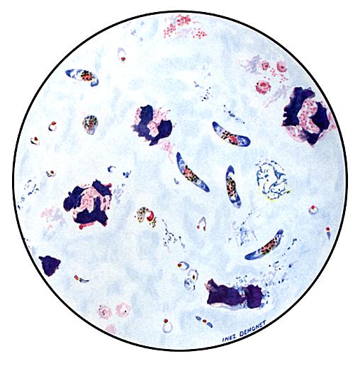
Thick Blood Smears
Illustrations from:
Wilcox A. Manual for the Microscopical Diagnosis of Malaria in Man.
Washington: U.S. Department of Health, Education and Welfare; 1960.
|
|