|
|
[Last Modified: ] |
|
|
|
| [Plasmodium
falciparum] [Plasmodium malariae] [Plasmodium ovale] [Plasmodium vivax] |
|
|
|
|
|
|
|
|
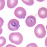
Plasmodium falciparum: Trophozoites
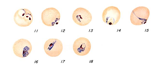 |
Figs. 11-18:
Increasingly mature trophozoites.
Illustrations from:
Coatney GR, Collins WE, Warren M, Contacos PG. The Primate Malarias. Bethesda: U.S.
Department of Health, Education and Welfare; 1971.
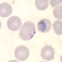
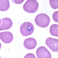
Smears from patients:
Although rarely seen in peripheral blood smears, older, ring stage parasites are referred to as trophozoites. The cytoplasm of mature trophozoites tends to be thicker and more dense than in younger rings. As P. falciparum trophozoites grow and mature, they tend to retain their ring-like shape and sometimes trace amounts of yellow pigment can be seen within the cytoplasm. Growing trophozoites in P. falciparum can appear slightly amoeboid in shape.
 |
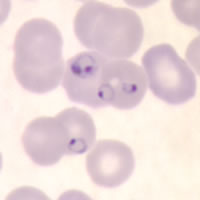 |
| A | B |
A: Appliqué
trophozoite. Note the slight amoeboid appearance of the parasite.
B: A
larger, more mature trophozoite and smaller, younger ring stage parasites
in a thin blood smear.
 |
 |
| C | D |
C, D: More mature and compact trophozoites. Note the presence of pigment in C.
|
|||