|
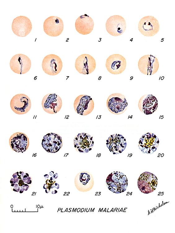
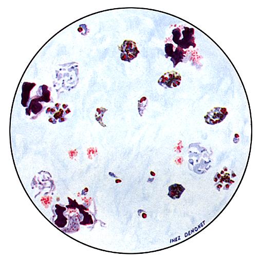
Plasmodium
malariae: Blood Stage Parasites
Thin Blood Smears
Fig. 1: Normal
red cell; Figs. 2-5: Young trophozoites (rings); Figs. 6-13: Trophozoites; Figs. 14-22: Schizonts; Fig. 23: Developing
gametocyte; Fig. 24: Macrogametocyte (female); Fig. 25:
Microgametocyte (male).
Illustration from:
Coatney GR, Collins WE, Warren M, Contacos PG. The Primate Malarias. Bethesda:
U.S. Department of Health, Education and Welfare; 1971.
Thick Blood Smears
Illustration from:
Wilcox A. Manual for the Microscopical Diagnosis of Malaria in Man.
Washington: U.S. Department of Health, Education and Welfare; 1960.
|