|
|
[Last Modified: ] |
|
|
|
| [Plasmodium
falciparum] [Plasmodium malariae] [Plasmodium ovale] [Plasmodium vivax] |
|
|
|
|
|
|
|
|
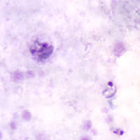
Plasmodium malariae: Ring Stage Parasites
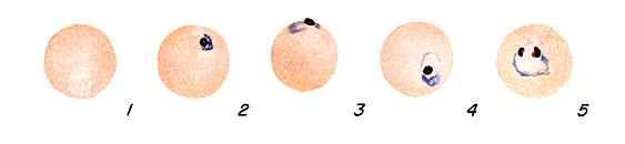 |
Fig. 1:
Normal red cell; Figs. 2-5: Rings.
Illustrations from:
Coatney GR, Collins WE, Warren M, Contacos PG. The Primate Malarias. Bethesda: U.S.
Department of Health, Education and Welfare; 1971.
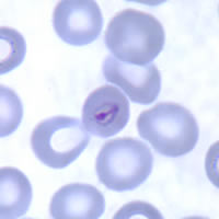
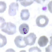
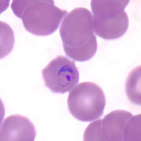
Smears from patients:
Plasmodium malariae rings have sturdy cytoplasm and a large chromatin dot. The red blood cells (RBCs) are normal to smaller than normal (3/4×) in size.
 |
 |
 |
| A | B | C |
A, B, C: Ring forms in thin blood smears.
 |
| D |
D: A thick blood smear showing two rings (lower right) and a gametocyte.
|
|||