|
|
[Last Modified: ] |
|
|
|
| [Plasmodium
falciparum] [Plasmodium malariae] [Plasmodium ovale] [Plasmodium vivax] |
|
|
|
|
|
|
|
|
Plasmodium ovale: Trophozoites
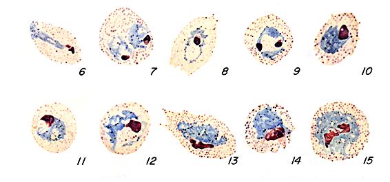 |
Increasingly mature
trophozoites. Note the fimbriated red cells (Figs. 8, 13).
Illustrations from: Coatney
GR, Collins WE, Warren M, Contacos PG. The Primate Malarias. Bethesda: U.S. Department
of Health, Education and Welfare; 1971.
Smears from patients:
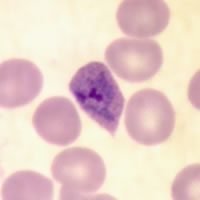
Plasmodium ovale trophozoites have sturdy cytoplasm, large chromatin dots, and can be compact to slightly amoeboid. Red blood cells are normal to slightly enlarged (1 1/4×), may be round to oval, and are sometimes fimbriated. Schüffner's dots are visible under optimal conditions.
 |
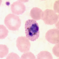 |
| A | B |
A, B: Trophozoites of P. ovale in thin blood smears. A is slightly amoeboid. B shows a more compact trophozoite and Schüffner's dots.
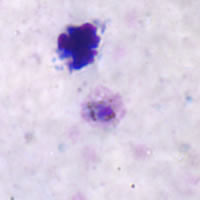 |
| C |
C: Trophozoite in a thick blood smear.
|
|||