|
|
[Last Modified: ] |
|
|
|
| [Plasmodium
falciparum] [Plasmodium malariae] [Plasmodium ovale] [Plasmodium vivax] |
|
|
|
|
|
|
|
|
Plasmodium vivax: Trophozoites
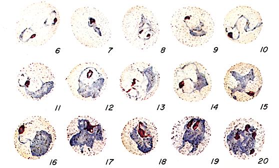 |
Figs. 8-18:
Increasingly mature trophozoites of P. vivax.
Illustrations from: Coatney GR, Collins WE, Warren M, Contacos PG. The Primate
Malarias. Bethesda: U.S. Department of Health, Education and Welfare; 1971.
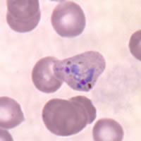
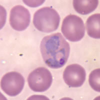
Smears from patients:
Plasmodium vivax trophozoites show amoeboid cytoplasm, large chromatin dots, and have fine, yellowish-brown pigment. Red blood cells are enlarged 1 1/2 to 2× and may be distorted. Under optimal conditions, Schüffner's dots may appear more fine than those seen in P. ovale.
 |
 |
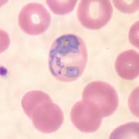 |
| A | B | C |
A, B, C: Large, amoeboid trophozoites of P. vivax in thin blood smears.
|
|||