|
|
[Last Modified: ] |
|
|
|
|
[Brachiola spp.] [Encephalitozoon
cuniculi] [Encephalitozoon hellem] [Encephalitozoon intestinalis (syn. Septata intestinalis)] [Enterocytozoon bieneusi] [Nosema spp.] [Pleistophora sp.] [Trachipleistophora spp.] [Vittaforma corneae (syn. Nosema corneum)] |
|
|
|
|
|
|
|
|
Transmission electron microscopy
 |
| A |
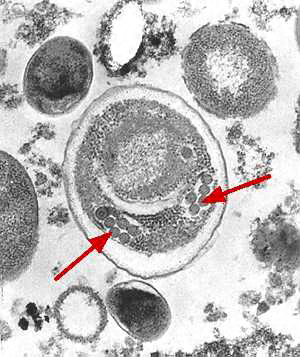
A: Electron micrograph of an Enterocytozoon bieneusi spore. Arrows indicate the double rows of polar tubule coils in cross-section which characterize a mature E. bieneusi spore.
 |
| B |
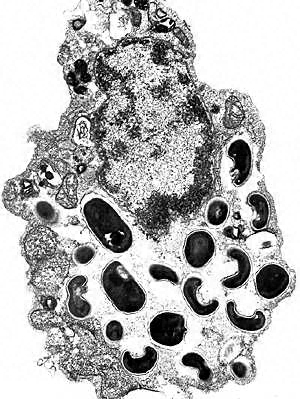
B: Electron micrograph of an eukaryotic cell with Encephalitozoon intestinalis spores and developing forms inside a septated parasitophorous vacuole. This is a characteristic feature of this microsporidian species.
|
|||