|
|
[Last Modified: ] |
|
|
|
|
[Brachiola spp.] [Encephalitozoon cuniculi] [Encephalitozoon
hellem] [Encephalitozoon intestinalis (syn. Septata intestinalis)] [Enterocytozoon bieneusi] [Nosema spp.] [Pleistophora sp.] [Trachipleistophora spp.] [Vittaforma corneae (syn. Nosema corneum)] |
|
|
|
|
|
|
|
|
Molecular
methods
The genus Encephalitozoon
contains three species related to human infections: E. hellem, E. cuniculi,
and E. intestinalis (syn. Septata intestinalis). The differentiation
between E. hellem and E. cuniculi cannot be achieved by morphological
analysis either using light or electron microscopy. Two additional microsporidian
species found in humans (Enterocytozoon bieneusi and Vittaforma corneae
[syn. Nosema corneum]) can be identified by experienced microscopists only.
Thus, molecular diagnosis with species-specific PCR primers is commonly believed to be the
gold standard for identification of microsporidian species. Our PCR
methods do not cross-amplify about 20 other microsporidian species tested at CDC's laboratory.
 |
| A |
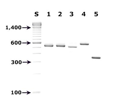
A: Agarose gel (2%) showing the diagnostic bands for species-specific PCR diagnostic primers designed for microsporidia that infect humans.
- Lane S: Molecular base pair standard (100-bp ladder). Black arrows show the size of standard bands.
- Lane 1: Encephalitozoon cuniculi positive specimen amplified with PCR primers ECUNF/ECUNR2, diagnostic band size: 549 bp.
- Lane 2: Encephalitozoon hellem template amplified with PCR primers EHELF/EHELR1, diagnostic band size: 547 bp.
- Lane 3: Encephalitozoon intestinalis template amplified with PCR primers SINTF1/SINTR3,4, diagnostic band size: 528 bp.
- Lane 4: Enterocytozoon bieneusi template amplified with PCR primers EBIEF1/EBIER15, diagnostic band size: 607 bp.
- Lane 5: Vittaforma cornae template amplified with PCR primers NCORF1/NCORR16, diagnostic band size: 375 bp.
References:
- Visvesvara GS, Leitch GJ, da Silva AJ, Croppo GP, Moura H, Wallace S, et al. Polyclonal and monoclonal antibody and PCR-amplified small-subunit rRNA identification of a microsporidian, Encephalitozoon hellem, isolated from an AIDS patient with disseminated infection. J Clin Microbiol 1994;32:2760-2768.
- De Groote MA, Visvesvara GS, Wilson ML, Pieniazek NJ, Slemenda SB, da Silva AJ, et al. Polymerase chain reaction and culture confirmation of disseminated Encephalitozoon cuniculi in patient with AIDS: Successful therapy with albendazole. J Infect Dis 1995;171:1375-1378.
- Visvesvara GS, da Silva AJ, Croppo GP, Pieniazek NJ, Leitch GJ, Ferguson D, et al. In vitro culture and serologic and molecular identification of Septata intestinalis isolated from urine of a patient with AIDS. J Clin Microbiol 1995;33:930-936.
- da Silva AJ, Slemenda SB, Visvesvara GS, Schwartz DA, Wilcox CM, Wallace S, Pieniazek NJ. Diagnosis of infections caused by the opportunistic microsporidian Septata intestinalis Cali et al. 1993 using PCR primers targeting the region coding for small subunit rRNA. Mol Diagn 1997 Mar;2:47-52.
- da Silva AJ, Bornay-Llinares FJ, del Aguila de la Puente C, Moura H, Peralta JM, Sobottka I, et al. Diagnosis of Enterocytozoon bieneusi (Microsporidia) infections by polymerase chain reaction in stool samples using primers based on the region coding for small-subunit ribosomal RNA. Arch Pathol Lab Med 1997;121:874-879.
- Pieniazek, NJ. unpublished.
|
|||