|
|
[Last Modified: ] |
|
|
|
|
[Brachiola spp.] [Encephalitozoon cuniculi] [Encephalitozoon
hellem] [Encephalitozoon intestinalis (syn. Septata intestinalis)] [Enterocytozoon bieneusi] [Nosema spp.] [Pleistophora sp.] [Trachipleistophora spp.] [Vittaforma corneae (syn. Nosema corneum)] |
|
|
|
|
|
|
|
|
Light
microscopy
Different
staining techniques can be used to demonstrate microsporidian spores in clinical samples.
The spores appear as oval to round shaped structures, measuring 1 to 4 µm (depending on the species). Some stains (Gram, Chromotrope 2R [A], and Quick-Hot Gram Chromotrope [B]) highlight a belt-like stripe in the equatorial diameter of the spores.
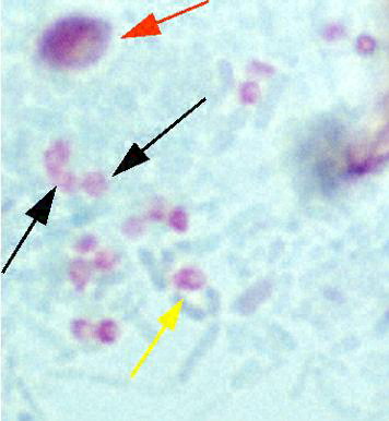 |
| A |
A: Stool smear stained with Chromotrope 2R containing Enterocytozoon bieneusi spores. Black arrows indicate E. bieneusi spores with their belt-like stripe accentuated. Red arrow indicates an unidentified yeast. The yellow arrow indicates a vacuolated spore.
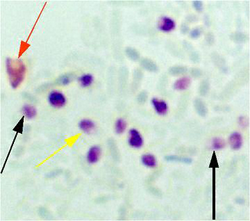 |
| B |
B: Stool smear stained with Quick-Hot Gram Chromotrope stain containing Enterocytozoon bieneusi spores. Black arrows indicate E. bieneusi spores with their belt-like stripe accentuated. The red arrow indicates an unidentified yeast. The yellow arrow indicates a vacuolated spore.
|
|||