|
|
[Last Modified: ] |
|
|
|
| [Schistosoma
mansoni] [Schistosoma haematobium] [Schistosoma japonicum] [Schistosoma mekongi] [Schistosoma intercalatum] |
|
|
|
|
|
|
|
|
Causal Agents:
Schistosomiasis is
caused by digenetic blood trematodes. The three main species infecting humans are Schistosoma
haematobium, S. japonicum, and S. mansoni. Two other species, more localized
geographically, are S. mekongi and S. intercalatum. In addition, other
species of schistosomes, which parasitize birds and mammals, can cause cercarial
dermatitis in humans.
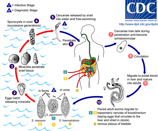
Eggs are eliminated with feces or urine
![]() .
Under optimal conditions the eggs hatch and
release miracidia
.
Under optimal conditions the eggs hatch and
release miracidia
![]() , which swim and penetrate specific snail intermediate
hosts
, which swim and penetrate specific snail intermediate
hosts
![]() .
The stages
in the snail include 2 generations of sporocysts
.
The stages
in the snail include 2 generations of sporocysts
![]() and the production of cercariae
and the production of cercariae
![]() . Upon
release from the snail, the infective cercariae swim, penetrate the skin of the human
host
. Upon
release from the snail, the infective cercariae swim, penetrate the skin of the human
host
![]() ,
and shed their forked tail, becoming schistosomulae
,
and shed their forked tail, becoming schistosomulae
![]() .
The schistosomulae migrate through several tissues and stages to their residence in the
veins (
.
The schistosomulae migrate through several tissues and stages to their residence in the
veins (![]() ,
,
![]() ).
Adult worms in humans reside in the mesenteric venules in various locations, which at times seem to be specific for each
species
).
Adult worms in humans reside in the mesenteric venules in various locations, which at times seem to be specific for each
species
![]() .
For instance, S. japonicum is more frequently found in the superior mesenteric
veins draining the small intestine
.
For instance, S. japonicum is more frequently found in the superior mesenteric
veins draining the small intestine
![]() ,
and S. mansoni occurs more often in the superior mesenteric veins
draining the large intestine
,
and S. mansoni occurs more often in the superior mesenteric veins
draining the large intestine
![]() .
However, both species can occupy either location, and they are capable of moving between
sites, so it is not possible to state unequivocally that one species only occurs in one location.
S. haematobium most often occurs in the venous plexus of bladder
.
However, both species can occupy either location, and they are capable of moving between
sites, so it is not possible to state unequivocally that one species only occurs in one location.
S. haematobium most often occurs in the venous plexus of bladder
![]() , but it can also be found in
the rectal
venules.
The females (size 7 to 20 mm; males slightly smaller) deposit eggs in the small venules of the portal and perivesical systems.
The eggs are moved progressively toward the lumen of the intestine (S. mansoni and S. japonicum) and of the bladder and ureters (S. haematobium), and are
eliminated with feces or urine, respectively
, but it can also be found in
the rectal
venules.
The females (size 7 to 20 mm; males slightly smaller) deposit eggs in the small venules of the portal and perivesical systems.
The eggs are moved progressively toward the lumen of the intestine (S. mansoni and S. japonicum) and of the bladder and ureters (S. haematobium), and are
eliminated with feces or urine, respectively
![]() . Pathology of
S. mansoni and S. japonicum schistosomiasis includes: Katayama fever,
hepatic perisinusoidal egg granulomas, Symmers’ pipe stem periportal fibrosis,
portal hypertension, and occasional embolic egg granulomas in brain or spinal cord.
Pathology of S. haematobium schistosomiasis includes: hematuria, scarring, calcification, squamous cell carcinoma, and occasional embolic egg granulomas in brain or spinal cord.
. Pathology of
S. mansoni and S. japonicum schistosomiasis includes: Katayama fever,
hepatic perisinusoidal egg granulomas, Symmers’ pipe stem periportal fibrosis,
portal hypertension, and occasional embolic egg granulomas in brain or spinal cord.
Pathology of S. haematobium schistosomiasis includes: hematuria, scarring, calcification, squamous cell carcinoma, and occasional embolic egg granulomas in brain or spinal cord.
Human contact with water is thus necessary for infection by schistosomes. Various animals, such as dogs, cats, rodents, pigs, hourse and goats, serve as reservoirs for S. japonicum, and dogs for S. mekongi.
Geographic
Distribution:
Schistosoma mansoni
is found in parts of South America and the Caribbean, Africa, and the Middle East;
S. haematobium in Africa and the Middle East; and S. japonicum in the Far East.
Schistosoma mekongi and S. intercalatum are found focally in Southeast Asia
and central West Africa, respectively.
|
||||||||