|
Microscopy
TAKE EXTREME
CARE IN PROCESSING THE SAMPLES!
INGESTION OF EGGS CAN RESULT IN CYSTICERCOSIS!
 |
 |
| A |
B |
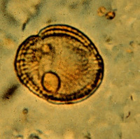
A, B: Taeniid eggs. The eggs of Taenia saginata
and Taenia solium are indistinguishable morphologically (morphologic
species identification will have to rely on the proglottids or scolices).
The eggs are rounded, diameter 31 to 43 µm, with a thick
radially striated brown shell. Inside each shell is an embryonated
oncosphere with 6 hooks. The egg in Figure B still has the primary membrane
that surrounds eggs in the proglottids.
 |
| C |
C: Pollen artifact that could be mistaken for a taeniid egg; however, the shell
is thinner, of nonuniform thickness, and no hooks are visible.
 |
 |
| D |
E |
 |
 |
| F |
G |
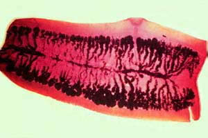
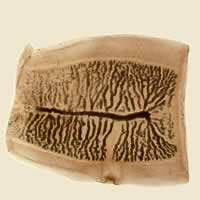
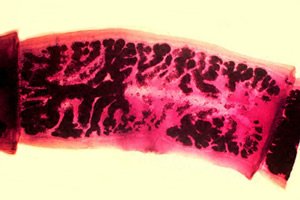
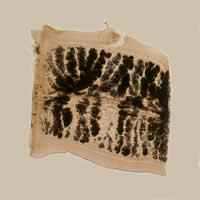
D, E, F, G:
Gravid proglottids of Taenia saginata (Figures D and E)
and Taenia solium (Figures F and G). Injection
of India ink in the uterus allows visualization of the primary lateral
branches. Their number allows differentiation between the
two species: T. saginata has 15 to 20 branches on each side
(Figure D and E), while Taenia solium has 7 to 13 (Figures
F and G). Note the genital pores in mid-lateral
position.
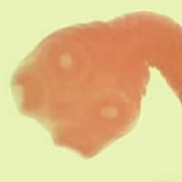
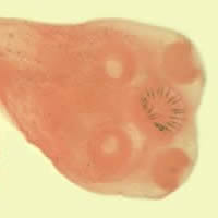
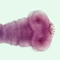
H, I, J:
Scoleces of Taenia saginata (Figure H) and Taenia solium
(Figures I and J). Scolex of T. saginata
has 4 suckers and no hooks. T. solium has 4 suckers in addition
to a double row of hooks.
 |
 |
| K |
L |
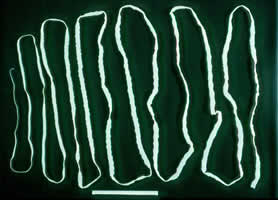
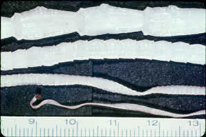
K, L: Taenia saginata adult worm. The adult in Figure K is 12
feet long.
|