|
|
[Last Modified: ] |
|
|
|
| [Toxoplasma gondii] |
|
|
|
|
|
|
|
|
Microscopy Findings
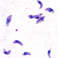 |
| A |
A: Toxoplasma gondii tachyzoites, stained with Giemsa, from a smear of peritoneal fluid obtained from a mouse inoculated with T. gondii. Tachyzoites are typically crescent shaped with a prominent, centrally placed nucleus.
 |
 |
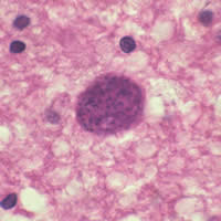
| B | C |
B: Toxoplasma
gondii in brain tissue stained with hematoxylin and eosin (100×).
C: Zoom of Image B, T. gondii cyst.
|
|||