|
 |
[Last Modified: ] |
 |
|
| [Entamoeba
histolytica] |
|
|
|
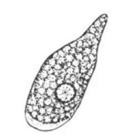
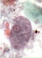
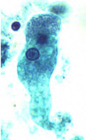
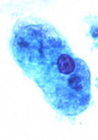
A, B,
C, D: Trophozoites
of Entamoeba histolytica/dispar, line drawing (A) and trichrome stain (B, C, and D).
(Reminder: in the absence
of erythrophagocytosis, the pathogenic E. histolytica is morphologically
indistinguishable from the nonpathogenic E. dispar!) Each trophozoite has a single nucleus, which has a centrally placed
karyosome and uniformly distributed peripheral chromatin. This typical
appearance of the nucleus is not always observed: some trophozoites can
have nuclei with an eccentric karyosome and unevenly distributed peripheral
chromatin. The cytoplasm has a granular or "ground-glass"
appearance. Entamoeba histolytica/dispar trophozoites
measure usually 15 to 20 µm (range 10 to 60 µm), tending to be more elongated
in diarrheal stool. B: Specimen contributed by Dr. Ray
Kaplan, SmithKline Beecham, Atlanta, GA.
|
|