|
|
[Last Modified: ] |
|
|
|
| [Entamoeba histolytica] | |
 |
 |
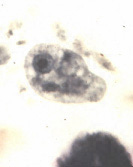 |
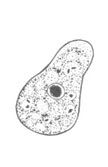
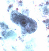
| A | B | C |
A, B, C: Iodamoeba buetschlii trophozoites. The trophozoites (line drawing, A; trichrome stain, B; iron hematoxylin stain, C) each have one nucleus with a large, usually central karyosome surrounded by refractile, achromatic granules. Their cytoplasm is coarsely granular, vacuolated and can contain bacteria, yeasts or other materials. The trophozoites measure usually 12 to 15 µm (range 8 to 20 µm).
|
||||||||