|
|
[Last Modified: ] |
|
|
|
| [Entamoeba histolytica] | |
 |
 |
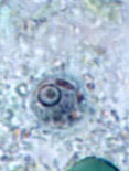 |
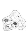
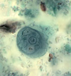
| A | B | C |
A, B, C: Entamoeba polecki trophozoites. The trophozoites (line drawing, A; trichrome stain, B and C) each have one nucleus that usually has small, discrete karyosomal chromatin and evenly distributed peripheral chromatin. Their cytoplasm is coarsely granular, vacuolated and can contain bacteria and yeasts (C). The trophozoites measure usually 15 to 20 µm (range 10 to 25 µm).
|
|||||||