|
 |
[Last Modified: ] |
 |
|
| [Entamoeba
histolytica] |
|
|
|
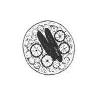
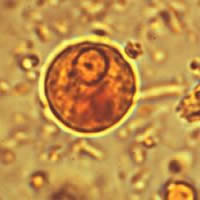
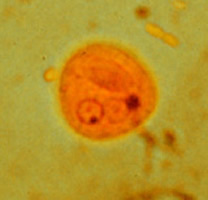
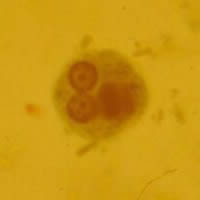
A, B,
C: Cysts of Entamoeba
histolytica/dispar, line drawing (A) and wet mounts stained
with iodine (B, C). The cysts are usually spherical
and often have a halo (B, C). Mature cysts have
4 nuclei. The cyst in B appears uninucleate while
in C, D, and E 2 to 3 nuclei are visible in the
focal plane (the fourth nucleus is coming into focus in
D). The nuclei have characteristically centrally located
karyosomes, and fine, uniformly distributed peripheral chromatin.
The cysts in C, D, and E contain chromatoid bodies
with the one in D being particularly well demonstrated,
with typically blunted ends. Entamoeba histolytica cysts
usually measure 12 to 15 µm.
 |
 |
|
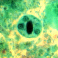
D |
E
|
D, E: Cysts of Entamoeba
histolytica/dispar, permanent preparations stained
with trichrome.
|
|