|
|
[Last Modified: ] |
|
|
|
|
[Brachiola spp.] [Encephalitozoon
cuniculi] [Encephalitozoon hellem] [Encephalitozoon intestinalis (syn. Septata intestinalis)] [Enterocytozoon bieneusi] [Nosema spp.] [Pleistophora sp.] [Trachipleistophora spp.] [Vittaforma corneae (syn. Nosema corneum)] |
|
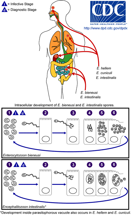 |
The infective form of microsporidia is the resistant spore and it can survive for a long time in the
environment
![]() . The spore extrudes its polar tubule and infects the host cell
. The spore extrudes its polar tubule and infects the host cell
![]() .
The spore injects the infective
sporoplasm into the eukaryotic host cell through the polar tubule
.
The spore injects the infective
sporoplasm into the eukaryotic host cell through the polar tubule
![]() .
Inside the cell, the sporoplasm
undergoes extensive multiplication either by merogony (binary fission) or schizogony
(multiple fission)
.
Inside the cell, the sporoplasm
undergoes extensive multiplication either by merogony (binary fission) or schizogony
(multiple fission)
![]() . This development can occur either in direct contact with the host cell
cytoplasm (e.g., E. bieneusi) or inside a vacuole termed parasitophorous vacuole
(e.g., E. intestinalis). Either free in the cytoplasm
or inside a parasitophorous vacuole, microsporidia develop by sporogony to mature
spores
. This development can occur either in direct contact with the host cell
cytoplasm (e.g., E. bieneusi) or inside a vacuole termed parasitophorous vacuole
(e.g., E. intestinalis). Either free in the cytoplasm
or inside a parasitophorous vacuole, microsporidia develop by sporogony to mature
spores
![]() . During sporogony, a thick wall is formed around the spore, which provides
resistance to adverse environmental conditions. When the spores increase in number and completely fill the host
cell cytoplasm, the cell membrane is disrupted and releases the spores to the
surroundings
. During sporogony, a thick wall is formed around the spore, which provides
resistance to adverse environmental conditions. When the spores increase in number and completely fill the host
cell cytoplasm, the cell membrane is disrupted and releases the spores to the
surroundings
![]() . These free mature spores can infect new cells thus continuing the
cycle.
. These free mature spores can infect new cells thus continuing the
cycle.
|
|||||||