| [Last Modified: ] | |
|
|
|
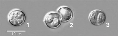
The image* below shows a wet preparation containing different phases of the Cyclospora cayetanensis oocyst visualized under differential interference contrast microscopy. Based on this image what is the correct statement?
(a)
The image demonstrates excystation of the C.
cayetanensis oocyst, from the unsporulated phase (image
1) to the sporulated phase, which contains two
sporocysts (image
3)
(b)
The image demonstrates sporulation of the C.
cayetanensis oocyst, from unsporulated phase (image 3)
to sporulated phase, which contains
two sporocysts (image 1)
(c)
The image demonstrates sporulation of the C.
cayetanensis oocyst, from unsporulated phase (image 1)
to sporulated phase, which contains two sporocysts (image
3)
(d)
All of the above
*image adapted from: Herwaldt BL. Cyclospora cayetanensis: a review, focusing on the outbreaks of cyclosporiasis in the 1990s. Clin Infect Dis 2000;31:1040-1057.
|
||||||||