|
Microscopy
Below are several Ascaris
eggs seen in wet mounts. Diagnostic characteristics:
- Fertilized eggs (A,
B on the right, D, F, G, H)
are rounded, have a thick shell, with an external mammillated layer that is often stained
brown by bile. In some cases, the outer layer is absent (decorticated eggs:
E, F on the right, G). Size: approximately 60 µm in diameter when spherical, and up to 75 µm when
ovoid.
- Unfertilized eggs (B
on the left, C, E) are elongated and larger (up to 90
µm in length); their shell is thinner; and their mammillated layer is more variable,
either with large protuberances (C) or practically none (E);
these eggs contain mainly a mass of refractile granules.
 |
 |
| A |
B |
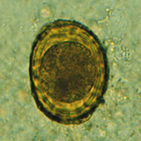
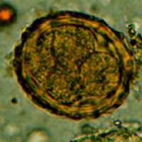
A:
Fertilized Ascaris
egg, still at the unicellular stage. Eggs are normally at this stage
when passed in the stool. Complete
development of the larva requires 18 days under favorable conditions.
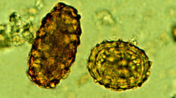
B: Unfertilized and fertilized eggs (left and right, respectively).
C: Unfertilized
egg. Prominent mammillations of outer layer.
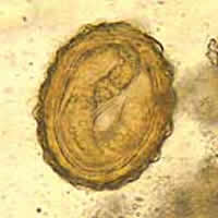
D: Fertilized egg. The embryo can be distinguished inside the egg.
E: Unfertilized egg with
no outer mammillated layer (decorticated).
 |
| F |
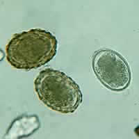
F:
Three fertilized eggs (one decorticated, on the right) of Ascaris
lumbricoides.
 |
 |
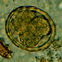
| G |
H |
G, H: Two fertilized eggs from the same patient, where embryos have begun to develop (this
happens when the stool sample is not processed for several days without refrigeration).
The embryos in early stage of division (4 to 6 cells) can be clearly seen.
Note that the egg
in G has a very thin mammillated outer layer.
 |
 |
| I |
J |
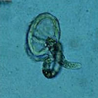
I: Egg
containing a larva, which will be infective if ingested.
J: Larva hatching from an egg.
Macroscopic (Gross) Observations
 |
| K |
K: An adult
Ascaris worm. Diagnostic characteristics: tapered ends; length
15 to 35 cm (the females tend to be the larger ones). This worm is a
female, as evidenced by the size and genital girdle (the dark circular
groove at bottom area of image).
|
|


