Answer
to Case 65
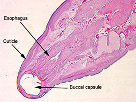
This was a case of hookworm infection. Several respondents thought this
was a trematode infection (e.g., Clonorchis spp., Paragonimus
spp., etc.). Tangential sections are not as good as cross-sections
to illustrate body morphology. However, in this case, the buccal
capsule is clearly visible. Several features excluded trematodes: the presence of a cuticle rather than tegument on the
body surface; a distinct layer of muscle cells composing the body wall;
and, most distinctly, the presence of a body cavity (best seen in Figure
C). Trematodes have a solid body composed of a parenchymatous
matrix in which the internal organs are embedded. Muscle fibers
course through the parenchyma, but do not form a heavy band as part of
the body wall as in nematodes.
Diagnostic features observed were:
- The worm possessed a body cavity.
- The large buccal capsule was clearly seen in Figures A and B.
- The size of the worm was within the range for hookworms (5 to 11 mm).
- The cuticle was thick.
- The esophagus was long and club-shaped and was clearly visible in all three images.
- The worms were located in the small intestine.
 |
| A |
Intact adult worms are usually required to differentiate the two primary species of hookworms infecting humans, Ancylostoma duodenale and Necator americanus. Morphological features such as the spicules found on male worms and presence of teeth or cutting plates (in male and female worms) have to be seen to make species identification. Due to the volume of international travel, the geographic distribution of both species has increased and become more overlapping, especially in southeast Asia. Historically, Ancylostoma duodenale was more prevalent in southern Europe, Sub-Saharan Africa, and Asia, whereas Necator americanus was typically found in the western hemisphere as well as southern Europe, southern Asia, India, Melanesia, and Polynesia.
For more information about hookworms, please click here.