Answer to
Case 9
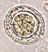
This specimen had a total of four parasite species:
- Figure A showed an Ascaris lumbricoides egg. The egg was partially decorticated, with only a few mammillations observable on the outer layer of the shell. It was fertilized, as evidenced by its thick shell, and its structured inner contents are separated from the shell by a space.
- Figure B showed another Ascaris lumbricoides egg. It was not fertilized. Compared to the fertilized egg (Figure A), it was more elongated, its shell was thinner, and its inner contents were not structured, but appear globular, and filled the shell, leaving no space. The imperfection on the upper right corner of the shell was somewhat misleading and suggested an operculum; however, careful examination showed that the shell was intact and that the outer covering was all that was missing.
- Figure C showed four Giardia intestinalis cysts, recognizable by their size, shape, and appearance. Since this wet mount preparation was not stained with iodine, the typical internal structures (axoneme, fibrils, nuclei) were not perfectly defined, but they were partially recognizable. You probably noticed the cysts in the background of the other lower magnification images—particularly in Figure D; even though they were out of focus, you may have guessed that they were Giardia cysts.
- Figure D showed a Trichuris trichiura egg. Straightforward diagnostic features included the typical barrel shape, the polar prominences or plugs, and the thick shell.
- Figure E showed an egg of Hymenolepis nana (dwarf tapeworm). Visible diagnostic features included: presence of hooklets (three of the six hooklets of the oncosphere can be seen), and size (50 micrometers by 44 micrometers, within the range for H. nana, which is 40 to 60 micrometers by 30 to 50 micrometers). The size distinguishes it from the larger H. diminuta (rat tapeworm) (70 to 86 micrometers by 60 to 80 micrometers). Another diagnostic feature, not observable in Figure E, would be the presence polar filaments between the oncosphere and the shell in H. nana. Such polar filaments are absent in H. diminuta. The image below (Figure A) shows these filaments (at the upper left and lower right corners) in an H. nana egg.
 |
| A |