|
Microscopy
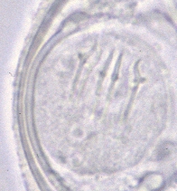 |
| A |
A: Egg of Dipylidium caninum.
The individual eggs are round to oval (average size 35 to 40 µm; range
31 to 50 µm by 27 to 48 µm) and contain an oncosphere that has 6
hooklets. Image contributed by the Oregon State Public Health
Laboratory.
 |
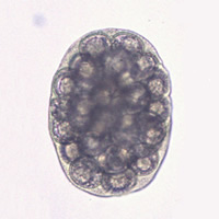 |
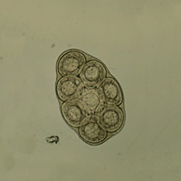
| B |
C |
B, C: Egg packets of Dipylidium caninum. Proglottids of
Dipylidium caninum contain characteristic egg packets
that are round to ovoid and contain 5 to 15 (sometimes more) eggs each. The egg packet
in B contains 8 visible eggs. The egg packet in C
(from the patient described in E) measures 230 µm
× 170 µm and contains many more eggs than the average number (over 30 eggs can be counted).
C: Image contributed by the Oregon State Public Health
Laboratory.
 |
 |
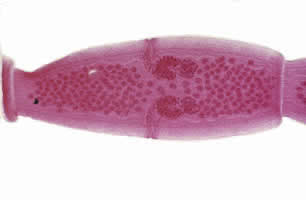
| D |
E |
D, E: Proglottids of Dipylidium
caninum. Such proglottids (average mature size 12 mm × 3 mm) have two genital
pores, one in the middle of each lateral margin. Proglottids may be passed singly or
in chains, and occasionally may be seen dangling from the anus. They are pumpkin
seed-shaped when passed and often resemble rice grains when dried. The genital pores
are clearly visible in the carmine-stained proglottid shown in D.
The proglottid shown in E (size 15 mm × 3 mm,
preserved in formalin), was passed by a 9 month old boy in the state of
Oregon (image contributed by the Oregon State Public Health Laboratory).
Macroscopic
(Gross) Observations
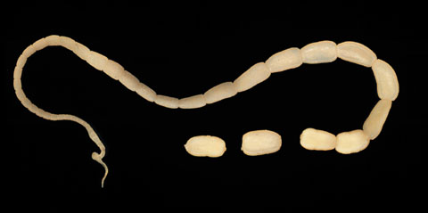 |
| F |
F: Adult tapeworm of
Dipylidium caninum. The scolex of the worm is very narrow and the proglottids, as they
mature, get larger.
|