|
Clinical Features:
The clinical
manifestations are localized but incapacitating. The worm emerges as a
whitish filament (duration of emergence: 1 to 3 weeks) in the center of a
painful ulcer, accompanied by inflammation and frequently by secondary
bacterial infection.
 |
 |
| A |
B |
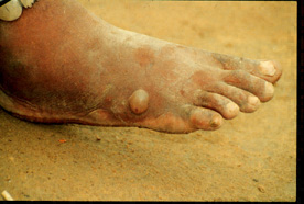
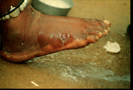
The female guinea worm induces a painful blister (A); after rupture of
the blister, the worm emerges as a whitish filament (B) in the center of
a painful ulcer which is often secondarily infected. Images contributed by Global
2000/The Carter Center, Atlanta, Georgia.
Laboratory
Diagnosis:
The clinical presentation of
dracunculiasis is so typical, and well known to the local population, that it does not
need laboratory confirmation. In addition, the disease occurs in areas where such
confirmation is unlikely to be available. Examination of the fluid discharged by the worm
can show rhabditiform larvae. No serologic test is available.
Treatment:
Local cleansing of the lesion and
local application of antibiotics, if indicated because of bacterial
superinfection. Mechanical, progressive extraction of the worm over a
period of several days. No curative antihelminthic treatment is
available.
|