|
|
[Last Modified: ] |
|
|
|
| [Brugia malayi] [Brugia timori] [Loa loa] [Mansonella ozzardi] [Mansonella perstans] [Mansonella streptocerca] [Onchocerca volvulus] [Wuchereria bancrofti] |
|
|
|
|
|
|
|
|
Microscopy (page 1 of 2)
 |
| A |
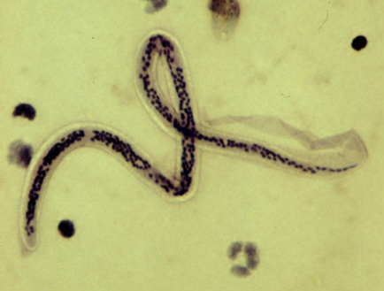
A: Microfilaria of Wuchereria bancrofti, from a patient seen in Haiti. Thick blood smears stained with hematoxylin. The microfilaria is sheathed, its body is gently curved, and the tail is tapered to a point. The nuclear column (the cells that constitute the body of the microfilaria) is loosely packed, the nuclei can be visualized individually and do not extend to the tip of the tail. The sheath is slightly stained with hematoxylin.
 |
| B |
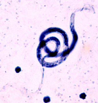
B: Microfilaria of Brugia malayi. Thick blood smear, hematoxylin stain. Like Wuchereria bancrofti, this species has a sheath (slightly stained in hematoxylin). Differently from Wuchereria, the microfilariae in this species are more tightly coiled, and the nuclear column is more tightly packed, preventing the visualization of individual cells.
 |
| C |
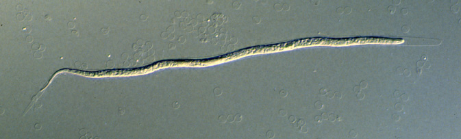
C: Microfilaria of Wuchereria bancrofti collected by filtration with a Nucleopore® membrane. Giemsa stain, which does not demonstrate the sheath of this sheathed species (hematoxylin stain will stain the sheath lightly). The pores of the membrane are visible.
 |
| D |
D: Microfilaria of Brugia malayi, collected by the Knott (centrifugation) concentration technique, in 2% formalin wet preparation. Note the erythrocyte ghosts (for size comparison). Note the clearly visible sheath that extends beyond the anterior and posterior ends of the microfilaria. There are four sheathed species: Wuchereria bancrofti, Brugia malayi, Brugia timori, and Loa loa.
 |
| E |
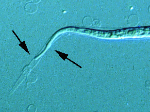
E: Detail from the microfilaria of Brugia malayi (see image above) showing the tapered tail, with a subterminal and a terminal nuclei (seen as swellings at the level of the arrows), separated by a gap without nuclei. This is characteristic of B. malayi.
|
||||||||