|
Microscopy
(page 2 of 2)
 |
| F |
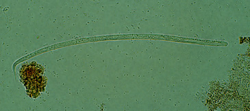
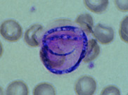
F: Microfilaria of
Onchocerca volvulus, from skin snip from a patient seen in
Guatemala. Wet preparation. Some important characteristics of the microfilariae of
this species are shown here: no sheath present; the tail is tapered and is sharply
angled at the end.
 |
| G |
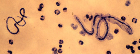
G: Microfilariae of
Loa loa (right) and Mansonella perstans (left).
Patient seen in Cameroon. Thick blood smear stained with hematoxylin.
Loa loa
is sheathed, with a relatively dense nuclear column; its tail tapers and is frequently
coiled, and nuclei extend to the end of the tail. Mansonella perstans is
smaller, has no sheath, and has a blunt tail with nuclei extending to the end of the tail.
 |
| H |
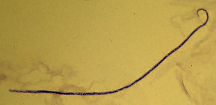
H: Microfilaria of Mansonella streptocerca from a skin snip.
Fixed in 2% formalin and stained with hematoxylin. The microfilaria is unsheathed, has a nearly straight body attitude, the tail is typically coiled into a “shepherd’s crook”, and terminal nuclei extend as a single row to the end of the tail.
 |
| I |
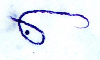
I: Microfilaria
of Mansonella ozzardi. Thick blood smear, stained with Giemsa. The
microfilaria is typically small, unsheathed, and has a slender, tapered tail that is
hooked ("button hook"). The nuclei do not extend to the end of the tail.
 |
| J |
J: An
artifact resembling a microfilaria. This is a mycelium of the fungus Helicosporium.
It can be differentiated from microfilariae by its small size (compare with the
erythrocytes in this thin shear), its characteristic shape and staining, and the absence
of regularly organized nuclei.
|