|
Microscopy
 |
| A |
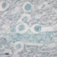
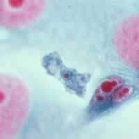
A: Naegleria fowleri trophozoites, cultured from cerebrospinal fluid. These cells
have characteristically large nuclei with a large, dark staining karyosome. The
amebae are very active and extend and retract pseudopods. Trichrome stain.
From a patient who died from primary amebic meningoencephalitis in Virginia.
 |
| B |
B: Naegleria
fowleri trophozoite in spinal fluid. Trichrome stain.
Note the typically large karyosome and the monopodial locomotion. Image
contributed by Texas State Health Department.
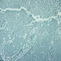
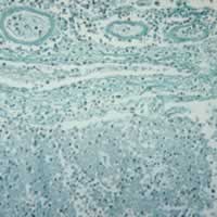
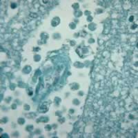
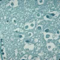
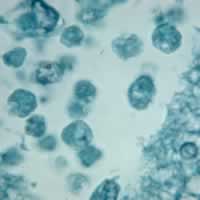
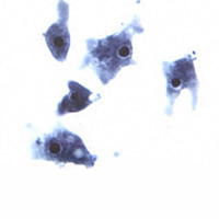
C,
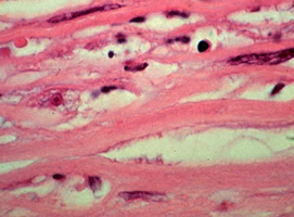
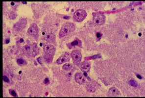
D, E, F, G, H: Naegleria fowleri in brain tissue, trichrome stain.
 |
 |
| I |
J |
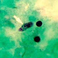
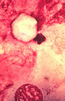
Acanthamoeba
sp. keratitis. I: Corneal biopsy showing a trophozoite; J: Cyst with the characteristic stellate shape,
in corneal scraping.
 |
| K |
K: Acanthamoeba
polyphaga trophozoite in tissue culture, trichrome stain.
 |
| L |
L: Balamuthia
mandrillaris trophozoites in brain tissue.
|