|
Microscopy
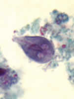
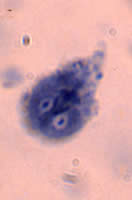
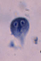
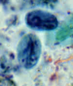
A, B, C: Three trophozoites
of Giardia intestinalis, stained with trichrome, (A), and
stained with iron hematoxylin
(B and C).
Each cell has two nuclei with a large, central karyosome. Cell size: 9
to 21 µm in length.
 |
| D |
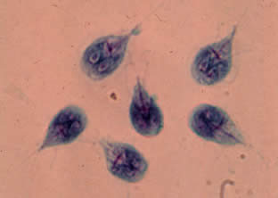
D: Giardia
intestinalis in culture. In these preparations, the flagellae (four pairs
per cell) are clearly visible.
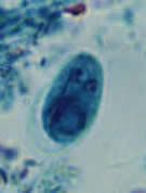
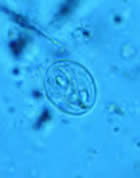
E, F, G:
Cysts of Giardia intestinalis, stained with iron hematoxylin (E, F)
and in a wet mount (G, from a patient seen in Haiti). Size:
8 to 12
µm in length. These cysts have two nuclei each (more mature ones will have four).
 |
| H |
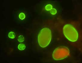
H:
Cysts of Giardia intestinalis (lower right) and oocysts of C. parvum
(upper left) labeled with commercially available immunofluorescent antibodies.
|
|