|
Microscopy
 |
 |
| A |
B |
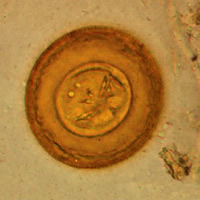
A: Egg
of Hymenolepis diminuta. These eggs are round or slightly oval, size 70
to 86 µm by 60 to 80 µm, with a striated outer membrane and a thin inner membrane. The
space between the membranes is smooth or faintly granular. The oncosphere has six
hooks (of which at least four are visible at this level of focus).
Image contributed by Georgia Department of Public Health.
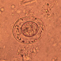
B: Egg
of Hymenolepis nana. These eggs are oval and smaller than
those of H. diminuta, their size being 40 to 60 µm by 30 to 50 µm. On the
inner membrane are two poles, from which 4 to 8 polar filaments spread out between the two
membranes. The oncosphere has six hooks (seen as dark lines at 8 o'clock).
Image contributed by Georgia Department of Public Health.
 |
| C |
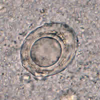
C: Artifact
resembling a H. nana egg. However, no hooks and no polar
filaments are visible in the artifact.
 |
 |
| D |
E |
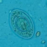
D: Hymenolepis
nana egg.
E: Hymenolepis
diminuta egg.
Macroscopic (gross) observations
 |
| F |
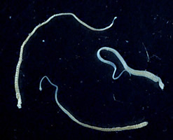
F: Three adult
Hymenolepis nana tapeworms. Each tapeworm (length: 15 to
40 mm) has a small, rounded scolex at the anterior end, and proglottids
can be distinguished at the posterior, wider end. Image
contributed by the Georgia Division of Public Health.
|