|
Microscopy
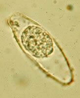
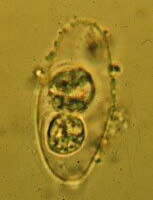
A, B,
C: Oocysts of Isospora belli.
The oocysts are large (25 to 30 µm) and have a typical ellipsoidal shape.
When excreted, they are immature and contain one sporoblast (A, B).
The oocyst matures after excretion: the single sporoblast divides in two sporoblasts (C),
which develop cyst walls, becoming sporocysts, which eventually contain four sporozoites
each. Images contributed by Georgia Division of Public Health.
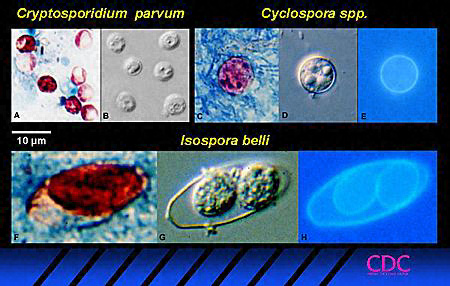 |
| Figure 2 |
Oocysts of Isospora belli
can also be stained with acid-fast stains, and can be visualized by epifluorescence on wet
mounts, as illustrated in Figure 2. Three coccidian parasites that most
commonly infect humans, seen in acid-fast stained smears (2A, 2C,
2F), bright-field differential interference contrast (2B,
2D, 2G) and epifluorescence (2E, 2H,
C. parvum oocysts do not autofluoresce).
|
|