|
Microscopy
 |
 |
| A |
B |
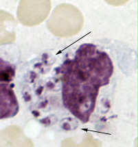
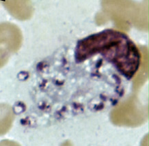
A, B: Leishmania tropica
amastigotes from an impression smear of a biopsy specimen from a skin
lesion. In A, an intact
macrophage is practically filled with amastigotes (arrows), several of which have
a clearly
visible nucleus and kinetoplast; in B, amastigotes are being
freed from a rupturing macrophage. Patient had traveled to Egypt, Africa,
and the Middle East. Based on culture in NNN medium, followed by isoenzyme analysis,
the species was L. tropica.
 |
| C |
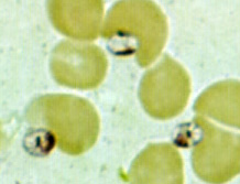
C: Three Leishmania
amastigotes, each with a clearly visible nucleus and kinetoplast, from the same impression smear as in A and B.
 |
| D |
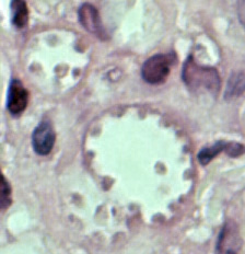
D: Leishmania mexicana
in a biopsy specimen from a skin lesion stained with hematoxylin and eosin.
The amastigotes are lining the walls of two vacuoles, a typical arrangement. The
species identification was derived from culture followed by isoenzyme analysis.
Infection was acquired in Texas.
|
|