|
 |
[Last Modified: ] |
 |
|
| [Entamoeba
histolytica] |
|
|
|
 |
 |
|
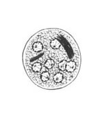
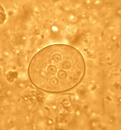
A |
B |
A, B: Cysts of Entamoeba
coli, line drawing (A), wet mount in iodine (B).
Mature cysts typically have 8 nuclei, and measure usually 15 to 25 µm
(range 10 to 35 µm). The cyst in B shows 5 nuclei visible in
this focal plane.
 |
 |
|
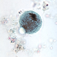
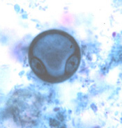
C |
D |
C, D: Cysts of Entamoeba
coli, trichrome stain. The cyst in D is an immature, binucleate form, frequently
seen with E. coli, where a large glycogen vacuole pushes the nuclei
to opposite sides. Chromatoid bodies are seen less frequently than
in E. histolytica. When present they are usually splinter
like with pointed ends (C) and thus different from the chromatoid
bodies of E. histolytica, which have rounded ends.
|
|