|
 |
[Last Modified: ] |
 |
|
| [Entamoeba
histolytica] |
|
|
|
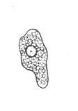
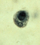
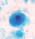
A, B,
C: Entamoeba hartmanni trophozoites, line drawing (A)
and trichrome stain (B and C). Entamoeba hartmanni
is often called a "small histolytica" because these two
species share many morphological characteristics, except their size.
The trophozoites of E. hartmanni (A, B, and C)
each have one nucleus with fine peripheral chromatin and a small, often
centrally located karyosome. The cytoplasm is finely granular. Note
that in B, the trophozoite has ingested a yeast, not
an erythrocyte. Ingestion of erythrocytes is pathognomonic of E.
histolytica. Trophozoites of E. hartmanni measure usually
8 to 10 µm (range 5 to 12 µm ) and are smaller than those of E. histolytica
(10 to 60 µm).
|
|