|
Microscopy
 |
 |
| A |
B |
 |
 |
| C |
D |
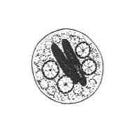
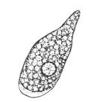
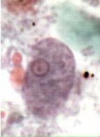
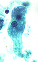
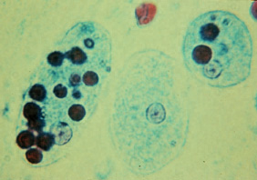
A, B,
C, D: Trophozoites of Entamoeba histolytica/dispar, line drawing (A) and trichrome stain (B,
C, D). Reminder: In the absence
of erythrophagocytosis, the pathogenic E. histolytica is morphologically
indistinguishable from the nonpathogenic E. dispar! Each trophozoite has a single nucleus, which has a centrally placed
karyosome and uniformly distributed peripheral chromatin. (This typical appearance
of the nucleus is not always observed: some trophozoites can have nuclei
with an eccentric karyosome and unevenly distributed peripheral chromatin.)
The cytoplasm has a granular or "ground-glass" appearance. Entamoeba
histolytica/dispar trophozoites measure usually 15 to 20 µm (range
10 to 60 µm), tending to be more elongated in diarrheal stool.
B: Specimen contributed by Dr. Ray Kaplan, SmithKline Beecham, Atlanta, GA.
 |
 |
| E |
F |
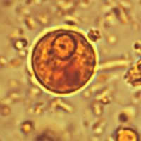
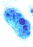
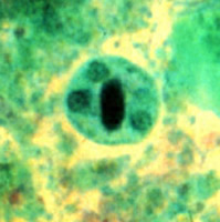
E,
F: Trophozoites of Entamoeba histolytica with ingested erythrocytes (trichrome stain).
The ingested erythrocytes appear as dark inclusions. Erythrophagocytosis
is the only morphologic characteristic that can be used to differentiate E. histolytica from the nonpathogenic
E. dispar. In these specimens, the parasite nuclei have the
typical small, centrally located karyosome, and thin, uniform peripheral
chromatin. E: Specimen contributed by the Texas
Department of Health.
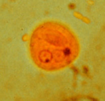
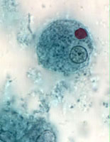
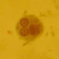
G, H,
I: Cysts of Entamoeba histolytica/dispar, line drawing (G), wet mounts stained with iodine (H,
I). The cysts are usually spherical and often have a halo (H,
I). Mature cysts have 4 nuclei. The cyst in H appears
uninucleate while in I, J, and K 2 to 3 nuclei are
visible in the focal plane (the fourth nucleus is coming
into focus in J). The nuclei have characteristically centrally
located karyosomes, and fine, uniformly distributed peripheral chromatin. The cysts in
I, J, and K
contain chromatoid bodies with the one in J being particularly well
demonstrated, with typically blunted ends.
Entamoeba histolytica cysts usually measure 12 to 15 µm.
 |
 |
| J |
K |
J, K: Cysts
of Entamoeba histolytica/dispar, permanent preparations stained with trichrome.
|
|