|
Microscopy
(page 2 of 3)
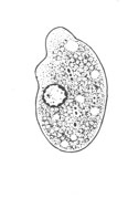
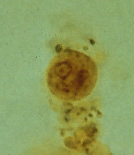
Nonpathogenic amebas (1)
A, B,
C: Trophozoites of Entamoeba coli, line drawing (A) and trichrome stain (B, C). The
trophozoites each have one nucleus with characteristically a large, eccentric
karyosome and coarse, irregular peripheral chromatin. The cytoplasm is
coarse and vacuolated ("dirty" cytoplasm) as illustrated in C.
Occasionally the cytoplasm contains ingested bacteria (B), yeasts or
other materials. The trophozoites of E. coli measure usually
20 to 25 µm, but they can be elongated (C) and reach up to 50 µm.
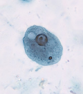
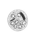
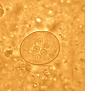
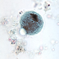
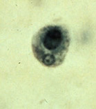
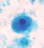
D, E,
F, G: Cysts of Entamoeba
coli, line drawing (D), wet mount in iodine (E),
and trichrome stain (F, G). Mature cysts
typically have 8 nuclei, and measure usually 15 to 25 µm (range 10 to
35 µm). The cyst in E shows 5 nuclei visible in this focal plane.
The cyst in G is an
immature, binucleate form, frequently seen with E. coli, where a large glycogen vacuole pushes the nuclei to opposite
sides. Chromatoid bodies are seen less frequently than in E.
histolytica. When present they are usually splinter like with
pointed ends (F) and thus different from the chromatoid bodies of E.
histolytica, which have rounded ends.
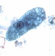
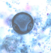
H, I,
J: Entamoeba hartmanni trophozoites, line drawing (H) and trichrome stain (I, J).
Entamoeba hartmanni is often called a "small histolytica"
because these two species share many morphological characteristics, except
their size. The trophozoites of E. hartmanni
(H, I, J) each have one nucleus with fine peripheral
chromatin and a small, often centrally located karyosome. The
cytoplasm is finely granular. Note that in I the trophozoite has ingested a
yeast, not an erythrocyte. (Ingestion of erythrocytes is pathognomonic of
E. histolytica.) Trophozoites of E. hartmanni measure usually
8 to 10 µm (range 5 to 12 µm ) and thus are smaller than those of E.
histolytica (10 to 60 µm).
 |
 |
| K |
L |
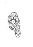
K, L: Entamoeba hartmanni
cysts, line drawing (K) and trichrome stain (L). Cysts
of E. hartmanni when mature have 4 nuclei and elongated chromatoid bodies with
rounded ends. Cysts of E. hartmanni measure usually 6 to 8
µm (range 5 to 10 µm) and thus are smaller than those of E.
histolytica (10 to 20 µm).
|
|