|
Microscopy
(page 3 of 3)
Nonpathogenic amebas* (2)
 |
 |
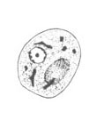
| A |
B |
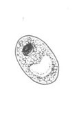
A,
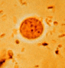
B: Endolimax nana trophozoites. The trophozoites each have one nucleus
with a characteristically large, irregularly shaped, blot-like karyosome
(line drawing, A; trichrome stain, B). Their nucleus has no peripheral
chromatin. Their cytoplasm is granular and vacuolated. The trophozoites
measure usually 8 to 10 µm (range 6 to 12 µm).
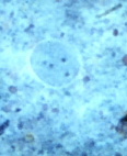
C, D,
E: Endolimax nana cysts. The cysts when
mature have 4 nuclei with large, blot-like karyosomes (line drawing,
C; wet mount in iodine, D; trichrome stain, E). The cysts of E. nana
do not have chromatoid bodies. The cysts measure 6 to 8 µm (range
5 to 10 µm).
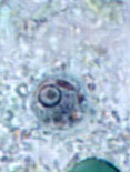
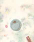
F, G,
H: Iodamoeba buetschlii trophozoites. The trophozoites (line
drawing, F; trichrome stain, G; iron hematoxylin stain,
H) each have one nucleus with a large, usually central karyosome surrounded by refractile,
achromatic granules. Their cytoplasm is coarsely granular, vacuolated
and can contain bacteria, yeasts or other materials. The trophozoites
measure usually 12 to 15 µm (range 8 to 20 µm).
 |
 |
| I |
J |
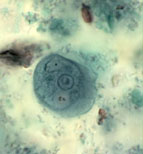
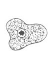
I,
J: Iodamoeba buetschlii cysts. The cyst (line
drawing, I; trichrome stain, J) have only one nucleus with a large, usually
eccentric karyosome. They do not have chromatoid bodies but have
a compact, well defined glycogen mass. The cysts measure usually
10 to 12 µm (range 5 to 20 µm) and their shape varies from ovoidal
to rounded.
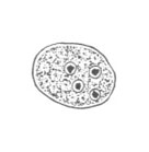
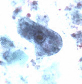
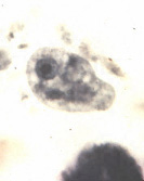
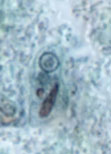
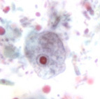
K, L,
M: Entamoeba polecki trophozoites. The trophozoites (line
drawing, K; trichrome stain, L and M ) each have one nucleus that usually
has small, discrete karyosomal chromatin and evenly distributed peripheral
chromatin. Their cytoplasm is coarsely granular, vacuolated and
can contain bacteria and yeasts (M). The trophozoites
measure usually 15 to 20 µm (range 10 to 25 µm).
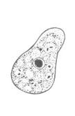
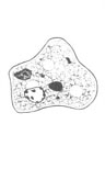
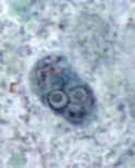
N, O,
P: Entamoeba polecki cysts. The cysts (line
drawing, N; trichrome stain, O and P) have one nucleus (rarely two,
P) with a small, usually eccentric karyosome (which can also be
rather pleomorphic). Their cytoplasm contains small inclusions and
an "inclusion mass", which stains only weakly in iodine.
The cysts measure usually 11 to 15 µm (range 9 to 18 µm) and their
shape varies from spherical to oval.
*The pathogenicity of Entamoeba polecki is currently unclear.
Further studies are necessary in order to satisfactorily resolve whether
E. polecki should be considered a pathogen or commensal.
|
|