|
|
[Last Modified: ] |
|
|
|
| [Schistosoma
mansoni] [Schistosoma haematobium] [Schistosoma japonicum] [Schistosoma mekongi] [Schistosoma intercalatum] |
|
|
|
|
|
|
|
|
Microscopy
Schistosoma haematobium
 |
 |
| N | O |
 |
 |
| P | Q |
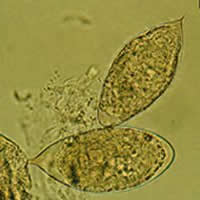
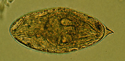
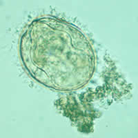
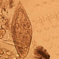
N, O, P, Q: Schistosoma haematobium eggs. In this species, the eggs are large and have a prominent terminal spine at the posterior end. Length 112 to 170 µm. In Figure O, the miracidium is shown inside the egg.
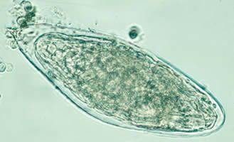
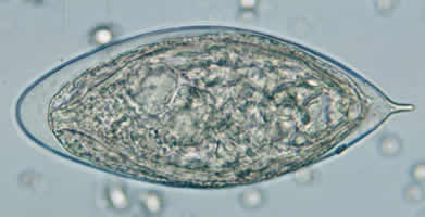
Schistosoma mekongi and Schistosoma intercalatum
 |
 |
| R | S |
R: Schistosoma
mekongi egg.
S: Schistosoma interculatum egg.
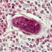
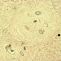
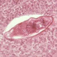
Cross-sections of human tissues with Schistosoma spp. eggs
 |
 |
 |
| T | U | V |
 |
 |
| W | X |
T, U,
V, W, X: Cross-section of different human tissues showing Schistosoma sp.
eggs.
T: Schistosoma mansoni eggs in intestinal wall.
U: Schistosoma japonicum eggs in colon
V: Schistosoma japonicum eggs in liver.
W, X: Schistosoma sp. in liver and bladder, respectively.
|
||||||||